The!Mad!Scientist!
  • Home
  • Courses
    • IB Chemistry Home Page
    • IB Biology Home Page
    • General Chemistry
  • G10 Science Home Page
  • Calendar

G11 SL IB Biology

Test Topics

12/14/2016

0 Comments

 
Magnification
Differences between and features of prokaryotes and eukaryotes
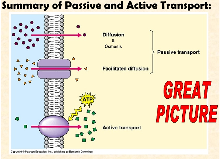
Diffusion and osmosis
Exceptions to cell theory; multiple nuclei
Cell theory
Liver cell micrograph (Google "liver cell micrograph" so you can get familiar with how they should look)
Route used to export proteins from the cell
Surface area/volume ratio
Identifying organelle structures in eukaryotes and prokaryotes
Facilitated diffusion requirements
Function of ribosomes in cells
Identifying structures of the plasma membrane
Draw and label an animal cell, a bacterium, and a plant cell.  Include organelles.  Be able to identify organelles by correct name.

0 Comments

December 12 - 22, 2016

12/12/2016

0 Comments

 
Housekeeping: Your exam covers 1.1-1.4.  I will quiz you over 1.5 & 1.6 in January before mocks.  The material for 1.5 and 1.6 will be posted before then if you want to get ahead.

Agenda: 
1.  Review 1.2
2.  Introduction to the plasma membrane

Content Review: 
Links:  1.1 Cell Theory   1.2 Ultrastructure  1.3 Membrane Structure 
Textbook Readings: Section 1.3 & 1.4

Student Missions:

Mission 1: In Da Club!!!
Mission Objectives.  You should be able to...
​1. 
Sketch and annotate a model of the plasma membrane.
2. Explain the purpose of cholesterol and proteins in the membrane.
What you should focus on is the role of proteins, cholesterol and the orientation of the membrane.  You should also be able to complete a basic sketch of the  plasma membrane and describe it using the following terms: phospholipid bilayer, amphipathic, phosphate head, fatty acid tail, hydrophilic, hydrophobic, polar, nonpolar, cholesterol function and protein functions.  Additional information about membrane structure is of course found here.

For the test, you will be asked to DRAW and annotate a diagram of the plasma membrane.

Mission 2: HIStory. *sigh* 
Mission Objectives.  You should be able to...
1. Describe the structure of the plasma membrane.
2. Compare and contrast the Davson-Danielli model with the Fluid Mosaic Model


As you know, nature of science is one of the tenets of the IB curriculum.  A component is understanding the dynamic nature of scientific knowledge and how what we know changes based on observable evidence.  

Davson-Danielli's Lipid Bilayer.
  Basically, in 1935, these scientists developed a model of the plasma membrane and called it the lipid bilayer.  Lipids, of course, are fats (and oils) and bilayer means "two layers."  Their model suggested that the membrane was constructed like an ice cream sandwich, with the lipid bilayer acting as the cream and being surrounded by globular proteins (aka the cookies).​
Picture
Singer & Nicolson's Fluid Mosaic.  In 1972, much of what was known about the then-accepted lipid bilayer model had to be discarded, as the model couldn't explain the lack of symmetry some membranes have, and the fact that a protein layer simply doesn't work because it is mostly non-polar and wouldn't interface with water, which is essential. This, along with other evidence, was gathered by electron microscopes and observing cell cultures in solutions. As a result, scientists Seymour Singer & Garth Nicolson came up with the currently accepted model of the plasma membrane, the Fluid Mosaic.

All cellular membranes, whether plasmic or organelle, have the same general structure.  It is a phospholipid bilayer.  A phospholipid has a phosphate head and a fatty acid tail.  The phosphate head is polar and hydrophilic, meaning it interfaces with water extremely well.  The fatty acid tail is nonpolar and hydrophobic, meaning it does not interface with water.  Similar in idea to the lipid bilayer, the Fluid Mosaic forms a sandwich with the phosphate heads on the outside and the fatty acid tails tucked in.  However, there are cholesterol and protein molecules embedded throughout the phospholipid bilayer, giving it a mosaic appearance (see p. 28 for a nice image).  
Mission 3:  Back Up In Da Club!!!
Mission Objectives.  You should be able to...
1.  Explain how substances move through the plasma membrane.
2.  Describe the processes involved in active and passive support.
3.  Compare and contrast endocytosis and exocytosis.
4.  Describe the function of the Na/K pump.


Let's talk about substance transportation into and out of the cell. The function of the plasma membrane is to maintain homeostasis by allowing substances and nutrients into and out of the cell.  Basically, it acts like a bouncer at Club Cell.  What processes are at work?  There are two main types of transport: passive and active. 

Terms to know:  diffusion, facilitated diffusion, osmosis, Na/K pump, endocytosis, and exocytosis.  What is the relationship of equilibrium to passive transport but not active transport?


Big Question:  How does the plasma membrane allow nutrients and water into and out of cell?  Is this an easy process or no?  Is there a difference between how substances enter and how they exit?  Are the substances that enter the same as the ones which exit?  
Cellular transport moves substances within the cell and into and out of the cell.  There are several processes at work here, grouped under two classes: passive and active.

Passive Transport

Diffusion: This is the net movement of particles from an area of high concentration to an area of low concentration.  Think of how scents and fragrances make their way to your nose.  If someone sprays perfume, the scent is strongest near the bottle, but in time, the scent particle will make their way to your nose.  At some point, there will be an even distribution of scent particles in the air. 


Facilitated diffusion:  When substances move through the plasma membrane via a water-filled transport protein (called a channel protein).  Channel proteins open and close to allow necessary substances to diffuse through the membrane.

Osmosis:  This is a form of specialized diffusion of water across a selectively permeable membrane.  Usually the water is mixed with a solute (sometimes sugar or salt).  The water can pass through the membrane but the solute cannot.  The water will diffuse through the membrane towards the side with the greater concentration of solute particles until dynamic equilibrium is reached (when the concentration of the solution is the same on both sides of the membrane).

​Here's a nice passive transport animation with a few practice questions.  Here is another one on membrane transport.

Active Transport
Energy (ATP) is required to move substances into and out of the cell.  The proteins embedded in the plasma membrane are positioned so that part lies inside the bilayer and parts outside the bilayer.  They must work against a concentration gradient.  This means instead of substances (usually ions) moving from areas of high concentration to areas of low concentration, the proteins must work in reverse: moving ions from low concentration to high concentration. 

The Sodium/Potassium pump
Animal cells have a higher concentration of potassium ions outside the cell whereas sodium ions are more concentrated inside the cell.  The cell maintains these conditions  by pumping K+ ions into the cell and pumping Na+ ions out of it.  A membrane protein is required for this to occur.  Here is a lovely animation showing how it works.  
Picture
Size and Charge:  How easily a substances can move across a membrane passively depends on two factors: size and charge.  Small, nonpolar substances will move easily across the membrane.  Substances that are polar (water excepted), large, or both, do not cross membranes easily.  Examples of substances that move with ease are oxygen, nitrogen and carbon dioxide.  Substances that do not move easily are ions (potassium, sodium and chloride), glucose, and sucrose.
​

Endocytosis is the active transport process by which a cell encloses a substance in a portion of the plasma membrane.  This process is dependent of the fluidity and the orientation of the plasma membrane to enclose particles or macromolecules to form a vesicle that then enters the cytoplasm of the cell.  The membrane then reattaches itself with the vesicle enclosed.

Exocytosis is the opposite process.  Materials are secreted or expelled from the cell at the plasma membrane. Protein exocytosis begins in the ribosomes of rough ER and progresses through a series of steps (p. 37-38) until the substance is secreted to the extracellular environment.  Both processes require ATP and are used for the transport of large particles.  Can you explain the differences between pinocytosis and phagocytosis?

Homework: Corresponding workbook pages.

0 Comments

Lab: Respiration

12/4/2016

0 Comments

 
Today we are going to do an enzyme lab. We will be looking at the effect of different substrates on glycolysis in yeast.

Title:  The Effect of DIfferent Substrates on Glycolysis in Yeast


Lesson Objectives: 
1.  To measure the amount of carbon dioxide produced using three different substrates.
2.  To estimate the rate of respiration in each case using data processing.
3.  To link the theory of respiration with our knowledge of carbohydrates.

Content Review: 
Glycolysis is the process by which glucose is converted into pyruvate. This ultimately forms pyruvic acid, which is used to supply energy to living cells.  More about glycolysis can be found on BioNinja.

Preparation is Key.  So today (12/7/16), we will spend part of the period prepping for your lab.  You need to clean and label  your test tubes.  I will provide hot water so that you can make sure they are clean.  Dry them with paper towel.  Use a metric ruler and measure 3 cm from the bottom of the tube and draw a line using a Sharpie.  Do this for 4 test tubes.  Number them 1, 2, 3 and 4.

Put your test tubes in the rack and using a post-it, write your names on your test tubes and put them in a safe space.  You will be given your bag of flour (which is 20 spatulas' worth), labeled cups, and a spatula to keep with your materials.  If you do this today, then you will be ready to begin the lab on the next day.

Read through the handout and form a hypothesis about what may happen and why. This is your ticket out the door for today.  You need to get it checked off by me, as it is going into the gradebook as a classwork assignment.

You will be working in groups of 4. Put all group members' names on the post-it.

On the handout is the template for the data you will collect.  We will be working with limited equipment, so we will do the best we can. You will have to share the electronic balances.

Procedure:
1.  Take 4 test tubes and measure 3cm from the bottom.  Mark it on the test tube with a Sharpie.  Number them 1 2 3 and 4.

2.  Label your cups like so:  1.  STARCH   2.  SUGAR   3.  GLUCOSE  4.  CONTROL
  For the 5th cup, label it MIXTURE, as you'll be mixing your yeast and flour in this cup.

3.  Measure 75mL of warm water using the graduated cylinder and pour it into the MIXTURE cup.  Add 4g of yeast to the MIXTURE cup.  Stir gently.

3.  Carefully pour all of the flour to the yeast/water mix and stir gently.  It will be somewhat loose.

4.  Split the dough into 4 equal portions using the spoons.  Put each portion into one of the labeled cups.

5.  Clean your spoon.

6.  To the "STARCH" cup, add two spatulas' worth of cornstarch. Stir gently

7.  To the "SUGAR" cup, add two spatulas' worth of sugar. Stir gently

8.  To the "GLUCOSE" cup, add two spatulas' worth of glucose. Stir gently

9.  DO NOT ADD ANYTHING TO THE "CONTROL" CUP!!

10. You and your partners should (at the same time) add 3cm of dough from the labeled cups to the matching numbered test tube and mark the height of the dough. Start timing. One person should be watching and timing one test tube.  For example, Hala should observe and time the STARCH tube while Lamya observes and times the SUGAR tube.

11.  Measure the height of the dough in the test tubes every 3 minutes starting at t = 0. Stop after 18 minutes, or 21 minutes if the dough rises slowly.  Time in minutes is your X-axis.  Dough height in centimeters is your Y-axis.

12.  While you wait, document the materials used and make notes on the uncertainties of the equipment (I'll tell you this part).  Also document any observations (color, smell, sound, condition of test tubes, etc.  This is your QUALITATIVE DATA, which goes before quantitatiave data in the lab report.

You will have to use hot water to wash the used test tubes.

When you are finished recording data, please clean up after yourselves.  Wash the test tubes and put them face down in the racks.  Throw away the excess dough and the used cups and spoons.  Wipe off the counters and make sure you don't leave anything on the floor.  Respect the classroom space, please.

Homework:  Bring your laptops to class on Sunday.  We will use Excel to create a table of your data, and then from the table, generate a line graph that shows time (X) versus dough height (Y).  You should have at least five X values and five corresponding Y values.  Your table should have a title and labeled axes. This will be included in your lab report.  You will have to determine if your hypothesis was supported or refuted and provide an explanation.

Begin working on your lab report.  The final report is due December 15. It is important to include all the elements of a lab report (you should know what they are by now), even safety precautions.  If you want me to give you feedback, give it to me before then.

0 Comments

    Archives

    March 2020
    January 2020
    December 2019
    October 2019
    September 2019
    August 2019
    May 2018
    April 2018
    February 2018
    January 2018
    November 2017
    October 2017
    September 2017
    August 2017
    April 2017
    March 2017
    February 2017
    January 2017
    December 2016
    November 2016
    October 2016
    September 2016
    June 2016

    RSS Feed

Proudly powered by Weebly