Mission 1: The Cell Cycle
Mission Objectives. You should be able to...
1. List and describe each phase in the cell cycle.
2. Describe how DNA forms chromosomes.
3. Describe and explain the role of cyclins during interphase.
4. Identify the phases of mitosis in electron micrographs.
5. Describe the purpose of and/or outcome of mitosis.
This should be nothing short of a comprehensive review of what you learned back in 9th grade. No shocked faces or faux outrage, ok???
Understandings |
Applications & Skills |
|
Mitosis is the splitting of the nucleus into two identical daughter nuclei
Chromosomes condense by supercoiling during mitosis Cytokinesis occurs after mitosis and is different in plant and animal cells Interphase is a very active phase of the cell cycle with many processes occurring in the nucleus and cytoplasm. Cyclins are involved in the control of the cell cycle Mutagens, oncogenes, and metastasis are involved in the development of primary and secondary tumors |
App: The correlation between smoking and cancer incidence
Skill: Identify the phases of mitosis in electron micrographs Skill: Determination of a mitotic index from a micrograph |
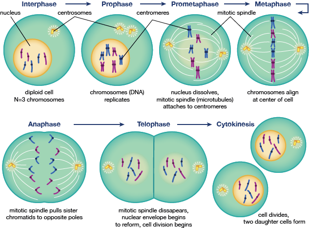
Image courtesy of shmoop.com.
The cell cycle describes the behavior of cells as they grow and divide. In most cases, the cell produces two cells that are genetically identical to the original. Cell division as a whole used to be called mitosis before more advanced cellular research determined that there are a number of significant nuclear processes that take place before the cell actually undergoes mitosis (called interphase). Mitosis itself has four phases: prophase, metaphase, anaphase, and telophase (PMAT).
The cell cycle describes the behavior of cells as they grow and divide. In most cases, the cell produces two cells that are genetically identical to the original. Cell division as a whole used to be called mitosis before more advanced cellular research determined that there are a number of significant nuclear processes that take place before the cell actually undergoes mitosis (called interphase). Mitosis itself has four phases: prophase, metaphase, anaphase, and telophase (PMAT).
|
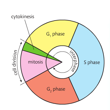
Interphase. This is the longest phase in the cell cycle. It is comprised of three smaller phases: G1, S and G2. In G1, the major event is cell growth. At this point, the cell is as small as it will ever be. In S, the major event is the replication of DNA. In G2, the cell actually grows and prepares for mitosis. Organelles increase in number and DNA condenses from chromatin to chromosomes and microtubules form. Special proteins called cyclins control the cell's progress through the cell cycle. Cyclins (there are two; one for each checkpoint) bond to substances called CDKs (cyclin-dependent protein kineases) that allow them to act as enzymes. Cyclins were discovered by accident. The first cyclin triggers DNA replication at the G1 checkpoint. The second cyclin triggers mitosis at the G2 checkpoint.
|
|
|
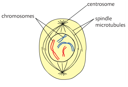
Prophase. During prophase (to the left), the nuclear envelope disappears and chromatin (loose DNA) coils to form chromosomes. Spindle fibers form and centrosomes move to the opposite ends of the cell.
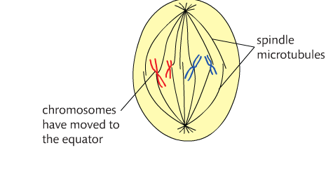
Metaphase. Chromosomes move to the center of the cell--called the metaphase plate (seen below). The centromeres lie on the equator of the plate. Each half of the chromosome is called sister chromatids. The spindle fibers move the chromosomes and the centrosomes are at opposite ends of the cell. |
|
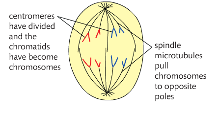
Anaphase. This is the shortest phase (seen below). The sister chromatids split and the chromosomes move towards opposite sides of the cell because the microtubules are shortening. Centromeres move first. At the end of this phase, each side (or pole) of the cell has an identical set of chromosomes.
|
|
|
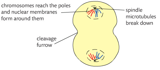
Telophase. The chromosomes are now at the poles of the cell and the nuclear envelope re-forms (seen below). The chromosomes uncoil to become chromatin once more. Nucleoli reappears, spindle fibers disappear, and the cell is now ready to undergo cytokinesis.
|
|
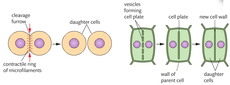
Cytokinesis. There is an inward pinching of the plasma membrane to form cleavage. In plant cells, due to the existence of the cell wall, a cell plate forms across the center of the cell. At the end of cytokinesis, there are two identical daughter cells. See below.
|
|
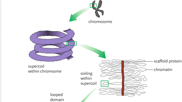
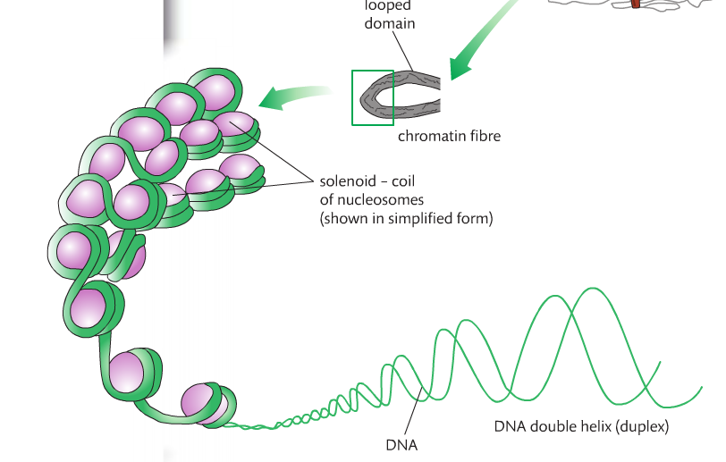
Chromatin condenses to form chromosomes through a process called supercoiling. The DNA wraps around histones (alkaline proteins ) to produce nucleosomes. Nucleosomes coil to form solenoids. Solenoids coil to form looped domains, and looped domains coil once more to form a chromosome.
|
|
Questions to consider: Why do you think interphase has to be divided into mini-phases? Can you describe what happens during each phase of mitosis? Can you SKETCH what happens during each phase of mitosis? Create a T-chart that describes each phase with a corresponding sketch.
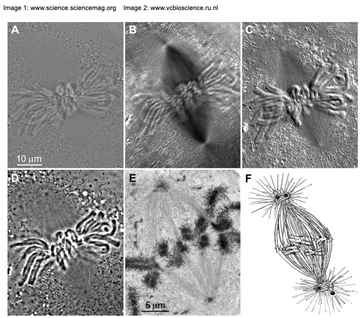
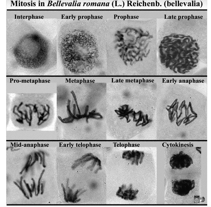
Can you identify the phases of mitosis in electron micrographs? Look at the examples below.
Can you identify the phases of mitosis in electron micrographs? Look at the examples below.
Mission 2: Cancer...Cells Gone Wild!
Mission Objectives. You should be able to...
1. Describe the development of primary and secondary tumors.
2. Explain the purpose and function of the mitotic index.
When mitosis goes awry, a mass of abnormal cells results, commonly called a tumor. A primary tumor is one that originates at the original site of a cancer. Secondary tumors, also known as metastasis, are tumors that spread from the primary site to another location in the body. Examples of metastasis would be a brain tumor composed of breast cancer cells or lung tumors composed of liver cancer cells. Metastasis can be so extensive that there may be multiple secondary tumors in different locations within the body.
Cancer is an insidious, nasty little killer...and there is still no known cure. The key to it may lie in one of your brains. I certainly hope so.
The Mitotic Index. The mitotic index (MI) is a ratio of cells in a tissue type undergoing mitosis to cells in the same tissue type not undergoing mitosis. A higher MI indicates rapid proliferation (to grow or multiply; specifically cells) of cells, and tumors with higher MIs may be harder to control and could lead to a poorer prognosis than a patient with a low MI. Practice determining the mitotic index with this virtual lab. This is the worksheet that goes with the lab.
Check out this creepy little horror movie showing normal cell growth versus cancerous cell growth. I needed a moment after watching this horrific thing.
The second video describes cells acting clean the funk up compared to cells who have home training. This video goes into significant detail about how cancerous cells form. Vocabulary you need to know: oncogenes, mutagens, mutation, mutagen and angiogenesis. Question to consider: How or why does a cancer cell forms?
Smoking is one of the leading causes of cancer. It is a mutagen. Cigarette corporations in the past have suppressed the results of tests that link smoking with cancer. It has only been within the past two or so decades that these corporations have had to acknowledge that smoking may lead to cancer and labels have been applied to cigarette packages and addenda to commercials added.
What do you think about the ethical considerations of corporations suppressing such results? Do you think that cigarette companies are the only corporations suppressing adverse data and/or results? What can you tell me about life, products, and companies that may engage in unethical practices in terms of reporting information so that citizens can make informed health decisions?
Homework: Corresponding workbook pages.